Tests and Diagnosis of Lung Cancer
Diagnosing lung cancer involves several types of tests and imaging. Generally an imaging is done first to identify the tumor then more invasive testing is used to confirm the diagnosis.
Finding lung cancer is fairly straightforward, once it is suspected. Diagnosing this disease is sometimes as easy as putting some sputum (spit) under the microscope. Lung cancer cells are often detected this way.
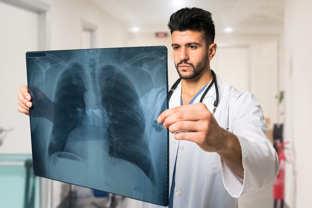
Other non-invasive tests for lung cancer include an x-ray and/or CT scan.

Your doctor may, however, decide that you need a biopsy which involves removing a small section of the lung to study it and detect abnormalities
The various diagnostic techniques associated with lung cancer are:
Imaging Tests for Lung Cancer:
Doctors generally perform imaging tests first to identify the cause of symptoms a patient might be experiencing or to gain a better look at a suspicious mass. There are five potential choices of imaging tests.
Doctors generally perform imaging tests first to identify the cause of symptoms a patient might be experiencing or to gain a better look at a suspicious mass. There are five potential choices of imaging tests.
Chest X-Ray: may be ordered to look for abnormal areas or masses in a lung.
CT Scan: may be ordered to obtain more detailed images of a certain area of the body that contains a suspicious mass. Tumors are more easily identified on CT scans than chest X rays.
MRI Scan: may be ordered to look for potential spread of lung cancer to areas like the brain or spinal cord.
PET Scan: may be ordered to identify fast growing tumors in various areas of the body and to identify if the cancer has spread to nearby lymph nodes.
Bone Scan: may be ordered to identify if the cancer has spread to the bones.
Diagnosis Tests for Lung Cancer:
Although an abnormal growth may be visible in certain imaging tests, diagnosis tests are performed to identify if the growth is cancerous. There are four types of diagnosis tests.
Sputum cytology: performed to identify if the sputum that is coughed up by a patient contains cancerous cells.
Thoracentesis: performed to identify if the fluid around the lungs contains cancerous cells.
Needle biopsy: performed in order to obtain tissue samples of a suspicious mass, growth, or tumor and identify if it contains cancerous cells.
Bronchoscopy: performed to identify potential growths present in the lung’s airway and biopsy them to determine if they contain cancerous cells.
Tests to Identify Lung Cancer spread:
These four tests are used to identify if the lung cancer has spread to distant tissues or organs. These tests are performed only if lung cancer has already been identified in the lungs.
These four tests are used to identify if the lung cancer has spread to distant tissues or organs. These tests are performed only if lung cancer has already been identified in the lungs.
Endobronchial ultrasound: performed to identify any distant lumps are present in areas like the lymph nodes. A biopsy can be performed on these areas to confirm whether or not they are cancerous.
Endoscopic esophageal ultrasound: performed to identify if there are any suspicious lumps present in the esophagus.
Mediastinoscopy and mediastinotomy: performed to identify if the areas between the lungs contain cancerous growths or cells.
Thoracoscopy: performed to identify if the cancer has spread to the chest wall.